Stem Cell Culture Media

Stem cells are characterized by self-renewal and multi-lineage differentiation potential. Under appropriate conditions, they can differentiate into various functional cell types. Mesenchymal stem cells (MSCs), in particular, do not elicit rejection in allogeneic transplantation, making them widely used in basic medical research and drug development. JSBio’s stem cell media are produced under GMP conditions, ensuring quality standards suitable for stem cell–based products. These formulations support efficient in vitro expansion of MSCs while maintaining multipotency.
Our Features

Our Strengths

Product Catalog

Product Catalog No. Form Package Size Description User Guide Inquiry and Trial
Stem Cell 1 11901-1492、11901-23049 Dry Powder、Liquid 500 mL、1000 mL、2 L、10 L、50 L、100 L、Customized Basal medium for stem cell culture and multipotency maintenance Download
Stem SFM 01 11903-25018 Liquid 500 mL Basal medium Download
Stem cell supplement1 99215-25021 Liquid 15 mL Supplement to Stem SFM 01 (with serum substitute) Download
Stem cell supplement2 99216-25022 Liquid 15 mL Supplement to Stem SFM 01 (growth factors only) Download

Frequently Asked Questio

No data

Case Studies

Stem Cell 1

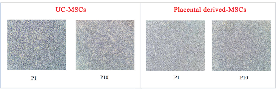
This medium promotes rapid MSC proliferation while maintaining stable morphology.

Figure 1. Morphology of umbilical cord–derived MSCs (UC-MSCs, left) and placenta-derived MSCs (PL-MSCs, right) cultured in Stem Cell 1, recorded using inverted microscopy.

Stem SFM 01

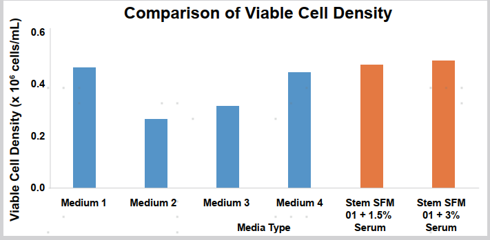
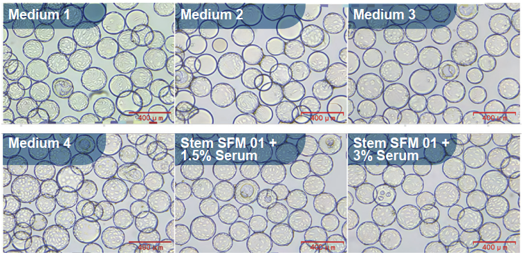
When culturing adipose-derived stem cells, Stem SFM 01 supplemented with only 1.5% fetal bovine serum produced cell morphology (Figure 2) and density (Figure 3) comparable to alpha-MEM supplemented with high concentrations of serum substitute, significantly reducing serum dependence and improving cost-effectiveness.

Figure 2. Morphology of adipose-derived stem cells on microcarriers.

Figure 3. Comparison of viable cell densities of adipose-derived stem cells.

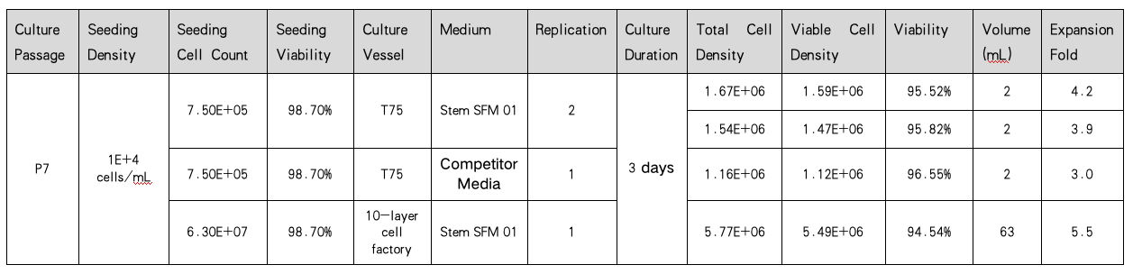
Table 1. Comparison of stem cell growth in Stem SFM 01 versus competitor media at passage 7. Stem SFM 01 showed superior viable cell density and fold expansion.

Surface Marker Analysis

Stem Cell 1

MSCs expanded in Stem Cell 1 while retaining high purity.


Table 2. Flow cytometric detection of surface markers of MSCs cultured to passage 5 (P5) in Stem Cell 1.

Stem SFM 01

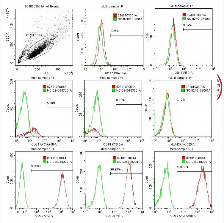
Figure 4. Positive marker expression exceeded 99% (CD73/CD90/CD105), while negative marker expression (CD34/CD45/HLA-DR) remained below 1%.

Multipotency Verification of MSCs

Stem Cell 1

Figure 5. Adipogenic and osteogenic induction experiments were performed using P5 UC-MSCs and PL-MSCs. Adipocytes were stained with neutral lipid dye (green), and osteoblasts were stained with alizarin red (red). MSCs at passage 5 retained multi-lineage differentiation potential.

Exosome Production

Stem SFM 01


Table 3. The exosome yield of Stem SFM 01 medium is 2.24×10¹² particles/L, higher than that of an MNC medium (2-5×10¹¹ particles/L).

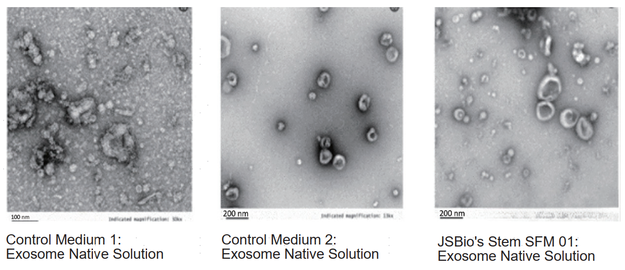
Figure 6. Comparison of exosome native solutions: exosomes produced in Stem SFM 01 exhibited superior productivity, along with uniform vesicle morphology and distinct structural features, indicating that Stem SFM 01 efficiently supports stem cells in secreting high-quality exosome